EYE (cont'd)
The Photoreceptors
There are two classes of photoreceptors in the human eye, although we will briefly mention a third photosensitive cell—figuring among ganglion cells—and its role in the section dedicated to the brain. These photoreceptors are the rods and cones.
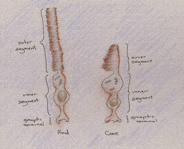
Rods are the more sensitive and the more numerous but are largely active in dark circumstance where greater sensitivity and number are needed. Cones function in lighted environments and are found, for the most part, in the fovea and the area immediately surrounding the fovea. They can be divided into subclasses according to which they are maximally sensitive: S-cones (short-wave), M-cones (medium-wave) and L-cones (long-wave). Somewhere, somehow along the brain-dominated portion of the visual pathway, these varying responses are associated with our notion of color.
Both rods and cones have the same structure: an inner segment composed of a synaptic body, a nucleus and the organelles required for ordinary cell metabolism. This segment is joined, by a ciliary stalk, to an outer segment, which is composed of thousands of disc membranes where tens of thousands of light-absorbing elements are found.
Of the photoreceptors in the eye, the rods are the most numerous. Though estimations vary—and on the order of millions—we will say that the retina of each eye contains some 120 million rods. In contrast, the cones may be as few as five million.
There are not, however, 125 million ganglion cells to convey their individual signals to the brain. If that were so, the area of the brain devoted to the processing of visual information—an area already substantially large—would then be of a size to compromise other cortical areas responsible for other sensory, motor and associative functions.
From an evolutionary perspective, this would result in an organism only able to live long enough to witness its own demise in an otherwise enviably detailed visual splendor.
Receptive Fields
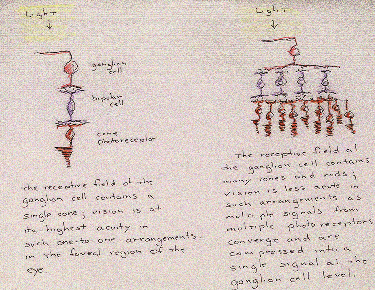
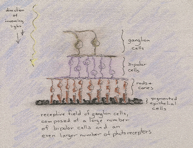
A ganglion cell will receive input from numerous bipolar cells, which will receive input, in turn, from even more numerous photoreceptors. In other words, a ganglion cell receives input, ultimately, from an entire field of rods or cones, or some combination thereof, constituting what we will now refer to as its receptive field.
A ganglion cell is a kind of representative body with a constituency of bipolar and photoreceptor cells; and just as a nation's population can be represented by a relatively tiny number of senators and congress people, so too can the 125 million or so rods and cones be represented by a substantially smaller one million ganglion cells.
The receptive field, incidentally, is arranged in a more or less circular pattern, having a center filled with excitatory or inhibitory cells around which is arranged a ring or surround of cells of an opposite, antagonistic character.
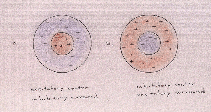
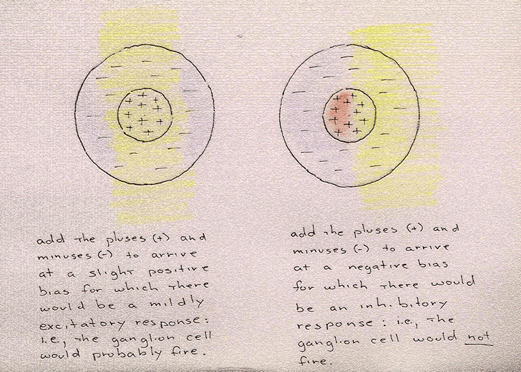
In figure A, light striking the center will beget an excitatory response in the ganglion cell; light striking the surround, an inhibitory one. But given the relative tininess of these receptive fields and the flood of light to which the retina is generally subjected, it is rare for but the center, or for but the surround, to be illuminated. So, when light strikes some or all of the center, and in some combination with the surround, the response is proportional to the combined number of excitatory and inhibitory responses. Where the numbers are equal, the ganglion cell—the ultimate register of these summated signals—will display little if any activity. Where excitatory signals outnumber inhibitory signals, the ganglion cell will fire, though not to the degree of frequency associated with maximum coverage of the center and minimum coverage of the surround.
Receptive fields, moreover, will vary in diameter. In the fovea, near the center of the eye, the receptive field of the ganglion cell will be very small, covering a very small number of photoreceptors—even as few as one. This is why vision is most acute, most fine, in this area. There is an almost one-to-one correspondence between the cones and the ganglion cells.
However, as we move out from the fovea, ganglion cells begin to receive signals from an ever-greater number of rods and cones in ever wider receptive fields: i.e., greater numbers of photoreceptors are represented by smaller numbers of ganglion cells with a resulting decrease in visual acuity.
